|
|
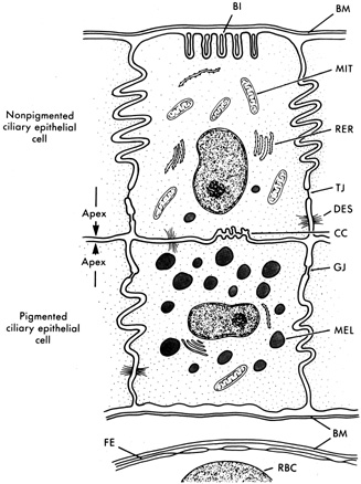
| Fig. 4. Schematic diagram of nonpigmented and pigmented epithelial cells. Note apices of cells facing each other. Basal infoldings (BI); basement membrane (BM); ciliary channels (CC); desmosomes (DES); fenestrated capillary endothelium (FE); gap junction (GJ); melanosome (MEL); mitochondrion (MIT); red blood cell (RBC); rough endoplasmic reticulum (RER); tight junction (TJ). (From Caprioli J: The ciliary epithelia and aqueous humor. In Hart M (ed): Adler's Physiology of The Eye, 9th ed, pp 228–247. St. Louis, Mosby Year-Book, 1992, with permission.) |