|
|
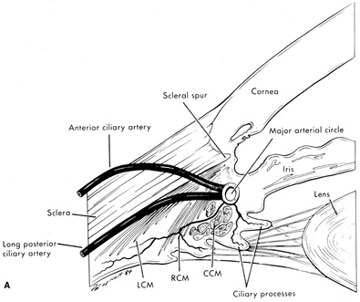
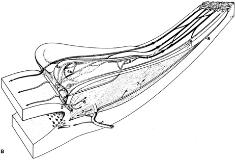
| Fig. 3. A: Blood supply to the ciliary processes. LCM, longitudinal ciliary muscle; RCM, radial ciliary muscle; CCM, circular ciliary muscle. B: Vascular architecture in the human ciliary body. (1), Perforating branches of the anterior ciliary arteries; (2), major arterial circle of the iris; (3), first vascular territory. The second vascular territory is depicted in 4a, marginal route and 4b, capillary network in the center of this territory. (5), third vascular territory; (6 and 7), arterioles to the ciliary muscle; (8) recurrent choroidal arteries. Light circles, terminal arterioles; dark circle, efferent venous segment. (A, From Caprioli J: The ciliary epithelia and aqueous humor. In Hart M, ed.: Adler's Physiology of the Eye, 9th ed. St. Louis: Mosby Year-Book, 1992:228–247, with permission; B, From Funk R, Rohen JW: Scanning electron microscopic study on the vasculature of the human anterior eye segment, especially with respect to the ciliary processes. Exp Eye Res 51:651, 1990, with permission.) |