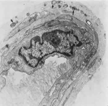
Fig. 19.
Electron micrograph of a human iridial arteriole from an older patient. Debris may be seen in the basement membrane surrounding the smooth muscle cells (
arrows
).