|
|
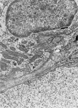
| Fig. 17. Electron micrograph of a portion of a capillary from the ciliary process facing the pigmented epithelium (p). Note the absence of an elastic lamina between the vessel and the pigmented epithelium. Fenestrae are present, and the endothelium is quite attenuated. This specimen was taken from a rat injected with thorium dioxide. l, capillary lumen. |