|
|
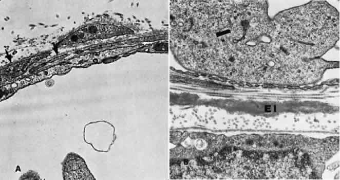
| Fig. 14. Electron micrograph. A. A portion of the capillary wall facing the suprachoroidea. The endothelial layer is relatively thick, and few fenestrae are present. Several pericytic processes (arrows) may be seen. B. A portion of the capillary wall facing the pigmented epithelium. Numerous fenestrae may be seen in the attenuated endothelium. The elastica of Bruch's membrane is well developed (EI). |