|
|
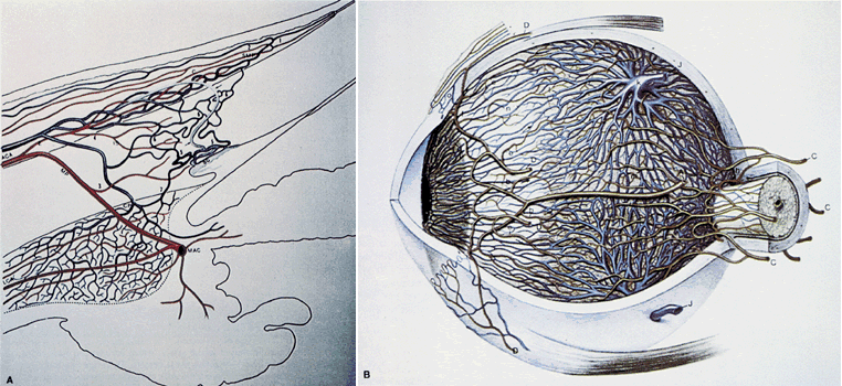
| Fig. 1. A. Drawing of a meridional section of the eye to show the blood supply of the limbal area. Red indicates arterial channels. An anterior ciliary artery (ACA) divides to form an episcleral (E) and a major perforating (MP) branch. The episcleral branches produce episcleral, conjunctival (C), and intra-scleral (IS) nutrient vessels. The conjunctival vessels form the superficial marginal plexus of the cornea (SMP). Two sets of vessels arise from the superficial marginal plexus: One (1) extends forward to form the peripheral corneal arcades; the other forms recurrent vessels (2) that run posteriorly to supply 3 to 6 mm of the perilimbal conjunctiva. The latter eventually anastomose with the recurrent conjunctival vessels from the fornices. The major perforating artery passes through the sclera to join the major arterial circle (MAC) of the iris. At 3, a branch from the major perforating artery passes forward to form the intrascleral arterial channels of the limbus. This region often is supplied by a vessel that arises directly from the anterior ciliary artery as an episcleral vessel, such as the one indicated at 4. Venous channels are blue. The major venous drainage from the limbus is into the episcleral veins, which then unite with the ophthalmic veins. The deep scleral venous plexus (5) is close to Schlemm's canal (SC). An aqueous vein (arrows) arises from the deep scleral plexus and joins the episcleral veins. The intrascleral venous plexus (6) forms an extensive network in the limbal stroma. An important part of the drainage from the ciliary plexus (CP) is into the deep and intrascleral venous plexus. One of these channels is seen at 7. B. The uveal blood vessels. The blood supply of the eye is derived from the ophthalmic artery. Except for the central retinal artery, which supplies the inner retina, almost the entire blood supply of the eye comes from the uveal vessels. There are two long posterior ciliary arteries, one entering the uvea nasally and on temporally along the horizontal meridian of the eye near the optic nerve (A). These two arteries give off three to five branches (b) at the ora serrata that pass directly back to form the anterior choriocapillaris. These capillaries nourish the retina from the equator forward. The short posterior ciliary arteries enter the choroid around the optic nerve (c). They divide rather rapidly to form the posterior choriocapillaris, which nourishes the retina as far anteriorly as the equator (the choriocapillaris is not shown in this drawing). This system of capillaries is continuous with those derived from the long posterior ciliary arteries. The anterior ciliary arteries (D) pass forward with the rectus muscles, then pierce the sclera to enter the ciliary body. Before joining the major circle of the iris, they give off 8 to 12 branches (e) that pass back through the ciliary muscle to join the anterior choriocapillaris. The major circle of the iris (f) lies in the corona ciliaris and sends branches posteriorly into the ciliary body as well as forward into the iris (g). The circle of Zinn (h) is formed by pial branches (i) as well as branches from the short posterior ciliary arteries. The circle lies in the sclera and furnishes part of the blood supply to the optic nerve and disc. The vortex veins exit from the eye through the posterior sclera (J) after forming an ampulla (k) near the internal sclera. Venous branches that join the anterior and posterior parts of the vortex system are meridionally oriented and are fairly straight (l), whereas those joining the vortices on their medial and lateral sides are oriented circularly about the eye (m). The venous return from the iris and ciliary body (n) is mainly posterior into the vortex system, but some veins cross the anterior sclera and limbus (o) to enter the episcleral system of veins. (Hogan MS, Alvarado J, Weddell J: Histology of the Human Eye. Philadelphia, WB Saunders, 1971) |