|
|
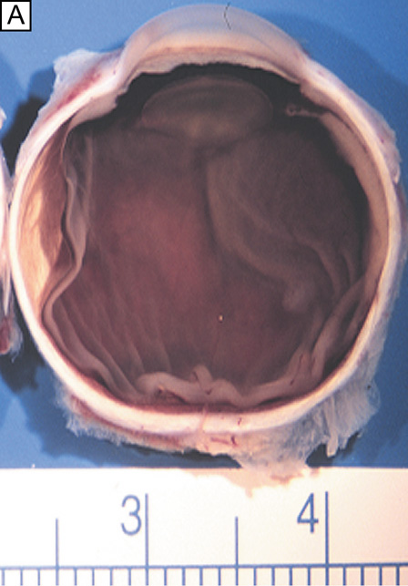
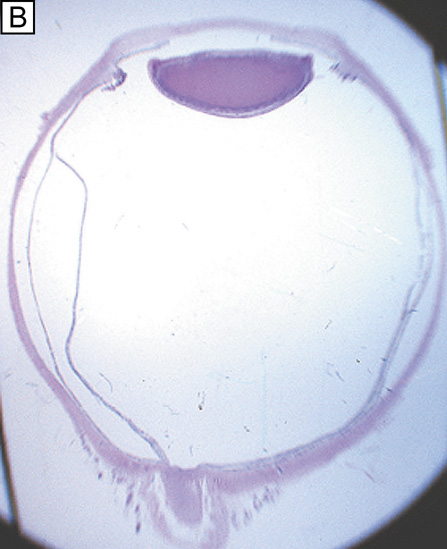
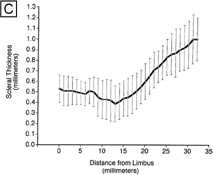
| Fig. 33. (A) Gross photo of the superior section of a horizontally bisected normal globe demonstrating the cross-sectional appearance of the sclera, limbus, and cornea. (B) Photomicrograph of a normal globe showing the cross-sectional appearance of the sclera, limbus, and cornea (PAS, 2X). (C) Line graph summarizing the average scleral thickness (± SD) vs. distance from the limbus in normal eyes (n = 550). ([C] is from Olsen TW, et al. Human sclera: thickness and surface area. Am J Ophthalmol 125:237, 1998.) |