|
|
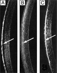
| Fig. 30. Slit lamp biomicroscopy of rabbit corneas 4 days (A) and 3 weeks (B) after manual epithelial debridement and 1 month after PRK (C). (A) demonstrates a 100 μm zone of keratocyte apoptosis and (B) demonstrates the repopulation of the acellular zone with migratory and activated keratocytes. Note that the epithelial debrided corneas completely returned to normal transparency by 8 weeks after injury. (C) demonstrates increased light scattering caused by myofibroblasts that are found in a hypercellular fibrotic scar. Note in the PRK corneas that the haze improved over the first year after surgery, but has not returned to normal levels. Arrows = area of interest. (Modified from Moller-Pedersen T. Keratocyte reflectivity and corneal haze. Exp Eye Res 78:553, 2004.) |