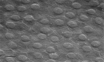
Fig. 23.
Scanning electron micrograph (1,000×) on the posterior surface of the corneal endothelium from a 65-year-old patient with healthy eyes. Note how the hexagonal endothelial cells form a uniform monolayer. Bar = 10 μm.