|
|
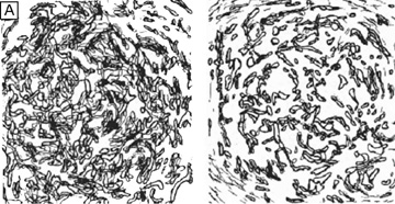
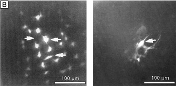
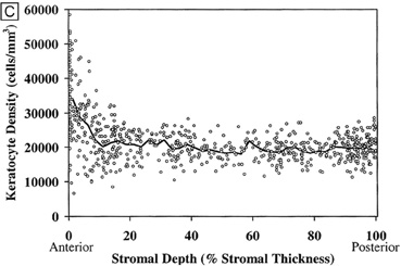
| Fig. 17. (A) Reconstruction of keratocyte outlines seen in frontal-section in the anterior and posterior thirds of the cellular corneal stroma. (Modified from Muller LJ, et al. Novel aspects of the ultrastructural organization of human corneal keratocytes. Invest Ophthalmol Vis Sci 36:2557, 1995.) (B) Fluorescent-dye spreading between many adjacent keratocytes in rabbit (center left) and human corneas (center right), which demonstrates the intimate importance of gap junctions in how keratocytes communicate with one another. (From Watsky MA. Keratocyte gap junctional dye spread in normal and wounded rabbit corneas and human corneas. Invest Ophthalmol Vis Sci 36:S22, 1995.) (C) Mean keratocyte density along the depth of central cellular corneal stroma. Notice the zone of increased density the closer one gets to the epithelial-stromal junction. Perhaps this is due to baseline, normal epithelial-stromal interactions. (From Patel SV, et al. Normal human keratocyte density and corneal thickness measurement by using confocal microscopy in vivo. Invest Ophthalmol Vis Sci 42:333, 2001.) |