|
|
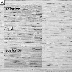
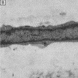
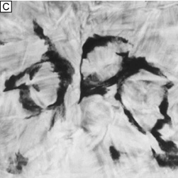
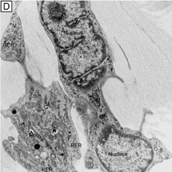
| Fig. 16. Light and TEM micrographs shown in cross and frontal-sectional views of the cellular corneal stroma demonstrating differences in the appearance of keratocytes depending on the cut, or perspective, of the section. (A) Cross-sectional light microscopy shows that keratocytes are primarily obliquely aligned to corneal surface in the anterior one-third of cellular corneal stroma and are aligned parallel to the corneal surface in the posterior two-thirds. (B) Cross-sectional TEM additionally shows that keratocyte nuclei occupy most of the area of the keratocyte seen in this perspective with only a thin rim of surrounding cytoplasm that contains only small numbers of cytoplasmic organelles. (C) From a tangential perspective, frontal-section light microscopy shows that keratocytes are arranged in a circular fashion. (D) Frontal-section TEM additionally shows that supposedly quiescent keratacytes may be more active in the baseline state than initially thought as an extensive amount of cytoplasmic organelles can be seen in this view. M, mitochondria; RER, rough endoplasmic reticulum; V, vacuoles. *Main portion of nucleus that contains nucleolus. (Modified from Muller LJ, et al. Novel aspects of the ultrastructural organization of human corneal keratocytes. Invest Ophthalmol Vis Sci 36:2557, 1995.) |