|
|
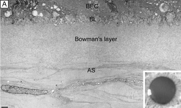
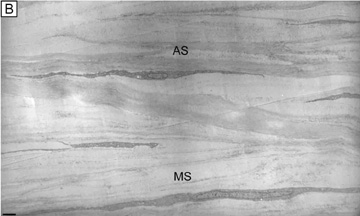
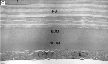
| Fig. 12. (A) The acellular Bowman's layer and anterior-most portion of the cellular corneal stroma. Notice that the interweaving lamellae of cellular stroma branch and insert into the posterior surface of the Bowman's layer forming an anterior corneal mosaic pattern after applying digital pressure on the corneal surface through the eyelid and fluorescein is instilled (inset). (B) The anterior third of the cellular corneal stroma showing the predominantly oblique lamellar orientation and the extensive vertical lamellar branches and interweaving. (C) The posterior third of the cellular corneal stroma, Descemet's membrane and endothelium. Notice the parallel oriented and predominantly orthogonal arrangement of lamellae in this portion of corneal stroma. Although some collagen fibrils randomly insert into Descemet's membrane, no specific pattern can be induced in this region of the cornea. BEC, basal epithelial cells; BL, basal lamina; AS, anterior stroma; MS, midstroma; PS, posterior stroma; BDM, banded portion of Descemet's membrane; NBDM, non-banded portion of Descemet's membrane; E, endothelial cells; ES, extracellular space. Bars = 1 μm. (TEM 4,750×.) |