|
|
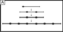
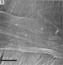
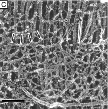
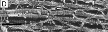
| Fig. 11. (A) Diagram of a type VI collagen molecule and how it assembles into filaments by aggregating into repeating tetramers of type VI molecules (periodicity of 100 nm). (B) Low magnification (6,200×) SEM showing bundles of collagen filaments extending between lamellae (arrow) and a loose meshwork of collagen filaments on the posterior surface of a corneal lamellae where an adjacent lamellae crosses it (arrowheads). Bar = 5 μm. (From Komai Y, et al. The three-dimensional organization of collagen fibrils in the human cornea and sclera. Invest Ophthalmol Vis Sci 32:2244, 1991.) (C) High magnification (115,000×) quick-freeze, deep-etched electron micrograph showing a loose meshwork of interlamellar beaded filaments with a periodicity of 100 nm (thick arrows) which appear to bind to collagen fibrils (long arrow) by their beads (arrowhead) and join fibrils from separate lamellae together. Bar = 0.2 μm. (D) Very high magnification (185,000×) quick-freeze, deep-etched electron micrograph of intralamellar collagen fibrils (long arrows) with beaded filaments (thick arrows) crisscrossing between fibrils and projecting three finger-like structures, which both appear to function in joining neighboring fibrils together. Bar = 0.1 μm. (C and D are from Hirsch M, et al. Three-dimensional supramolecular organization of the extracellular matrix in human and rabbit corneal stroma, as revealed by ultrarapid-freezing and deep-etching methods. Exp Eye Res 72:123, 2001.) |