|
|
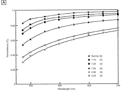
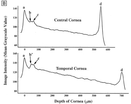
| Fig. 8. (A) Experimental values for the fraction of light transmitted through normal and edematous rabbit corneas as a function of wavelength. The ratio of the thickness of the edematous corneas to normal thickness values and the number of corneas used for each curve are given in the key. (From Farrell RA, et al. Wave-length dependencies of light scattering in normal and cold swollen rabbit corneas and their structural implications. J Physiol233:589, 1973.) (B) In vivo confocal microscopy back-scattered light intensity profiles from the central and temporal portions of a 25-year-old human cornea. Intensity peaks correspond to the (a) epithelium, (b) subbasal nerve plexus, (c) most anterior keratocytes layer, and (d) endothelium. (From Patel SV, et al. Normal human keratocyte density and corneal thickness measurement by using confocal microscopy in vivo. Invest Ophthalmol Vis Sci 42:333, 2001.) |