|
|
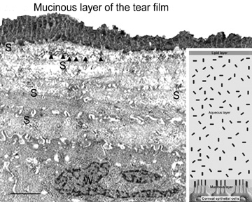
| Fig. 5 Transmission electron micrograph (10,000×) of surface epithelium from a specimen specially-preserved (glutaraldehyde + cetylpyridium chloride) and stained (tannic acid) to show the mucinous layer of the tear film (glycocalyx + membrane-bound mucins). Inset is a summary diagram showing how the tear film layers interact with the microvillae of the surface squamous epithelial cells. S, squamous cells; W, wing cells. Arrowheads = zonula occludens tight junctions. Arrowheads = zonula adherens tight junctions. Bar = 1 μm. |