|
|
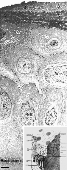
| Fig. 3. Transmission electron micrograph (3,500×) of the central corneal epithelium with a summary diagram (inset). Microvilli project from the anterior corneal surface into the tear film. S, squamous cells; W, wing cells; B, basal epithelial cells. Bar = 1 μm. (Inset modified from Hogan MJ, et al. Histology of the human eye. Philadelphia, WB Saunders, 1971.) |