|
|
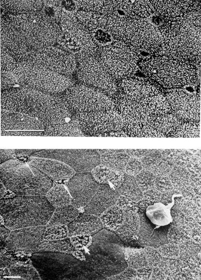
| Fig. 5. Electron micrographs of a conjunctiva. Top: Micrograph showing the conjunctiva with a uniform papillary appearance. Bar = 10 μm. Bottom: Micrograph showing the cell boundaries of the conjunctiva. Some cell boundaries are round (arrowhead). Small intercellular crypt openings are present (arrows). Mucin-like material is also present (white structure). Bar = 5 μm. (Reprinted from Greiner JV, Covington HI, Allansmith MR: Surface morphology of the human upper tarsal conjunctiva. Am J Ophthalmol 83:892–905, 1977.) |