|
|
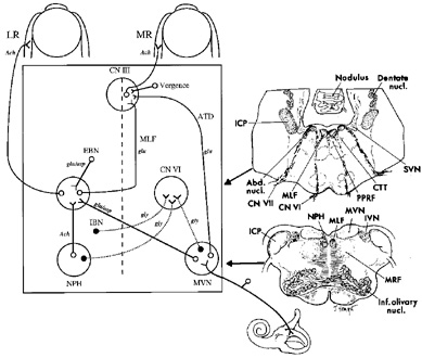
| Fig. 2. Anatomic scheme for the synthesis of signals for horizontal eye movements. The abducens nucleus (CN VI) contains abducens motoneurons that innervate the ipsilateral lateral rectus muscle (LR), and abducens internuclear neurons that send an ascending projection in the contralateral medial longitudinal fasciculus (MLF) to contact medial rectus (MR) motoneurons in the contralateral third nerve nucleus (CN III). From the horizontal semicircular canal, primary afferents on the vestibular nerve project mainly to the medial vestibular nucleus (MVN), where they synapse and then send an excitatory connection to the contralateral abducens nucleus and an inhibitory projection to the ipsilateral abducens nucleus. Saccadic inputs reach the abducens nucleus from ipsilateral excitatory burst neurons (EBN) and contralateral inhibitory burst neurons (IBN). Eye position information (the output of the neural integrator) reaches the abducens nucleus from neurons within the nucleus prepositus hypoglossi (NPH) and adjacent MVN. The medial rectus motoneurons in CN III also receive a command for vergence eye movements. Putative neurotransmitters for each pathway are shown: Ach, acetylcholine; asp, aspartate; glu, glutamate; gly, glycine. The anatomic sections on the right correspond to the level of the arrow heads on the schematic on the left. Abd. nucl., abducens nucleus; CN VI, abducens nerve; CN VII, facial nerve; CTT, central tegmental tract; ICP, inferior cerebellar peduncle; IVN, inferior vestibular nucleus; Inf. olivary nucl., inferior olivary nucleus; MVN, medial vestibular nucleus; MRF, medullary reticular formation; SVN, superior vestibular nucleus. (Modified from Leigh RJ, Zee DS. The Neurology of Eye Movements, 3rd ed. New York: Oxford University Press, 1999) |