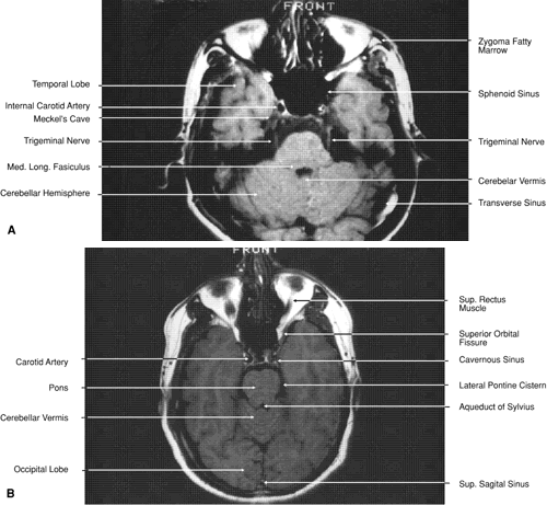
Fig. 30.
A.
Axial T1-weighted image at the level of floor of orbit and trigeminal nerve.
B.
Axial T1-weighted image at the level of oculomotor nerve.