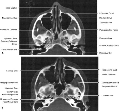
Fig. 29.
A.
Axial computed tomography soft tissue image at the level of the base of skull.
B.
Axial computed tomography bone window image at the level of the base of skull.