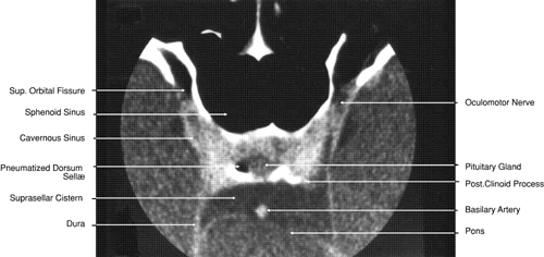
Fig. 28.
Axial computed tomography image with contrast medium through cavernous sinus and pituitary gland.