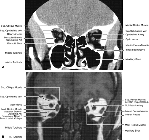
Fig. 19.
Coronal images through midorbit posterior to the globe.
A.
Computed tomography scan.
B.
T1-weighted magnetic resonance imaging.