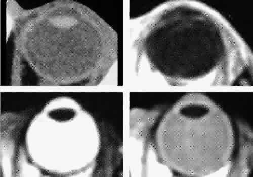
Fig. 12.
Axial cuts through the eye. Computed tomography (upper left), T1-weighted magnetic resonance imaging (upper right), T2-weighted magnetic resonance imaging (lower left), proton-density magnetic resonance imaging (lower right).