|
|
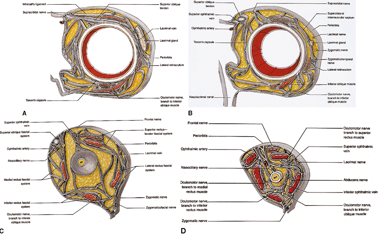
| Fig. 22. Fascial network of the orbit, oblique coronal views. A. Section through the equator of the globe. Note the diffuse attachments from the lateral rectus muscle sheath to the lateral retinaculum, collectively called the lateral rectus check ligament. Also shown are the complex attachments between each oblique muscle and its corresponding rectus muscle neighbor. B. Section through the posterior globe. The superior rectus-levator complex is clearly shown, with attachments to the orbital roof. C. Section at the lamina cribrosa. The superior ophthalmic vein begins to course more laterally, toward the superior rectus-levator complex. Attachments of the medial and lateral rectus muscle sheaths to their corresponding orbital walls are indicated. The lateral rectus attachments continue along the entire length of the lateral orbital wall. Note that the intraconal fat lies in discrete lobules separated by fascial sleeves. The orbital veins are contained within these sleeves, whereas the orbital arteries follow an independent course through the fat lobules. D. Section through the midorbit (anterior portion of the optic nerve). The superior ophthalmic vein is now shown in its fascial sling below the superior rectus muscle. More posteriorly, the orbital fascial system deteriorates, with a less defined intraconal space. (Dutton JJ: Atlas of Clinical and Surgical Orbital Anatomy, pp 103–105. Philadelphia, WB Saunders, 1994) |