|
|
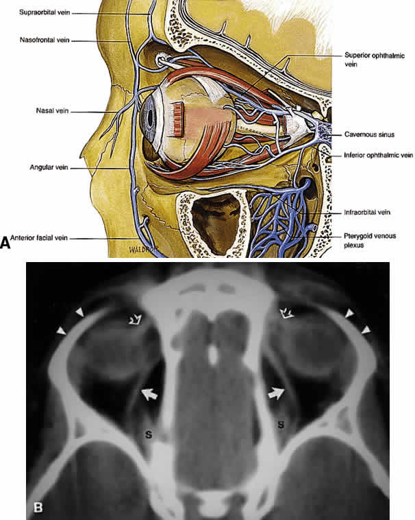
| Fig. 21. A. Venous drainage of the orbit. Major drainage is supplied by the superior ophthalmic vein, which drains into the CS. Note that the central retinal vein usually drains directly into the CS. Drainage through the pterygoid plexus is minor under normal conditions but becomes very important during outflow problems through the CS (carotid-cavernous or dural-cavernous fistulas). B. Axial CT reveals the course of the superior ophthalmic veins (closed arrows) beneath the superior rectus muscles (S). Also imaged are the superior orbital rims (arrowheads) and the superior oblique muscles (open arrows) as each passes through the trochlea. (A from Dutton JJ: Atlas of Clinical and Surgical Orbital Anatomy, p 79. Philadelphia, WB Saunders, 1994) |