|
|
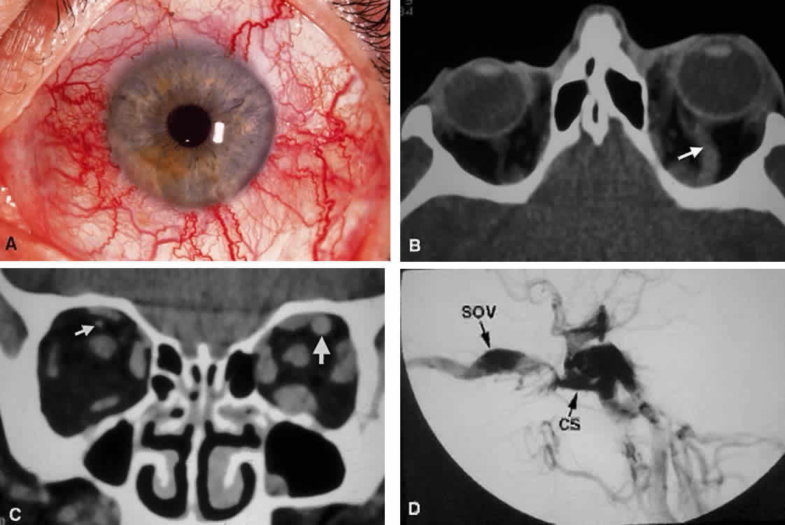
| Fig. 18. Carotid-cavernous fistula. A. Clinical photograph demonstrating tortuosity of the arteriolized episcleral veins, extending up to the limbus (the vascular congestion of conjunctivitis usually ends 1 mm short of the limbus). B. Axial CT shows the difference in caliber between the uninvolved superior ophthalmic vein and the involved vein (arrows). C. Coronal image likewise shows the difference in venous size (arrows). Also note the enlargement of the extraocular muscle on the involved side, indicative of orbital congestion. The superior ophthalmic vein is always found beneath the superior rectus muscle, to which it is tethered by a hammock-like fascial slip. On the involved side, orbital congestion and enlargement of the vein cause distortion of this anatomic relation. D. Parasagittal arteriography image of a different patient shows abnormal arterial filling of the CS, extending anteriorly into the orbit through an engorged superior ophthalmic vein (SOV). |