|
|
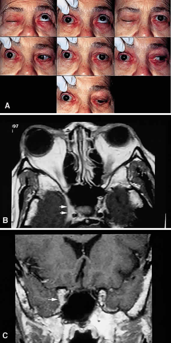
| Fig. 15. Parasellar syndrome. A. Composite photograph of an elderly patient with periorbital pain in the distribution of the right supraorbital nerve along with complete ptosis and absent levator function (upper left and upper right). When the eyelid was lifted, she complained of diplopia (center). Note the complete external ophthalmoplegia (remaining panels). MRI revealed a mass eroding the anterior clinoid process and extending into the CS and surrounding brain parenchyma. Given the bony erosion, a diagnosis of Tolosa-Hunt would be inappropriate in this case. A transorbital craniotomy for biopsy revealed metastatic adenocarcinoma. A systemic workup failed to reveal a primary site of involvement. The patient' orbital signs responded to radiation therapy. B and C. Axial and coronal MRI of a different patient who presented in an identical fashion. Note the enlargement of the right CS (arrows). Systemic workup was negative, and the patient responded rapidly to intravenous corticosteroids. The lesion disappeared on subsequent scans with no evidence of recurrence after 2 years. A diagnosis of “presumed Tolosa-Hunt syndrome”is acceptable in this case. |