|
|
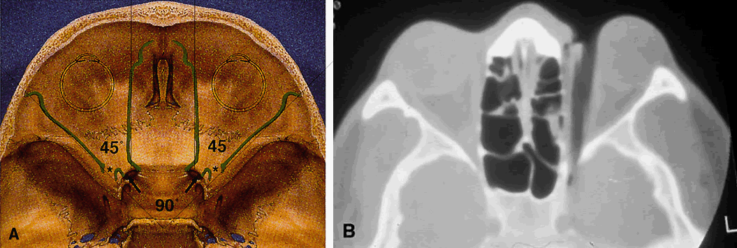
| Fig. 11. Osteology. A. An axial view of the orbital roof demonstrates the parallel course of the medial orbital walls (green). The lateral orbital walls (green) lie at an angle of 90° from each other, or 45° from each medial wall. Remember that the superior orbital fissure, and not the medially placed optic canal, lies at the posterior aspect of the orbit. B. CT of a wooden foreign body within the orbit after trauma. Note that the tip has traveled through the superior orbital fissure and lies within the CS, not the optic canal. In this case, the greatest worry was not the patient's vision, but the possibility of lacerating injury of the carotid siphon, which was confirmed on subsequent arteriography (A modified from Zide BM, Jelks GW: Surgical Anatomy of the Orbit, p 9. New York, Raven Press, 1985. B courtesy John W. Shore, MD, Austin, TX) |