|
|
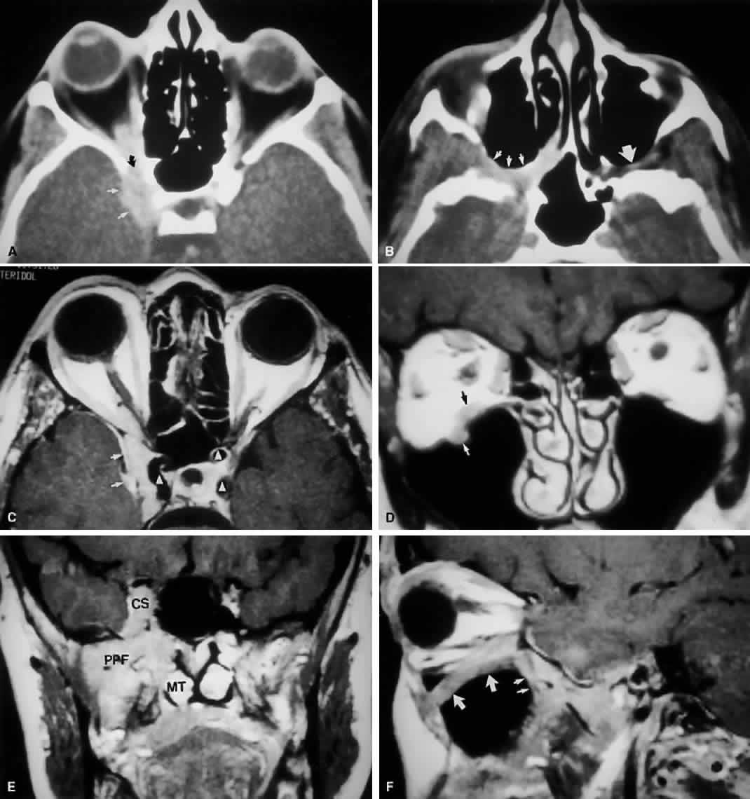
| Fig. 9. An orbital lymphoma involving the skull base provides accentuation of the apical spaces of the orbit. A. On this axial CT, the lesion infiltrates the CS, causing bulging and local invasion of its lateral dural wall (small arrows). Invasion into the orbital apex through the superior orbital fissure (curved arrow) is seen. Note that the patient is slightly rotated in the scanner, because the anterior clinoid and optic canal are visualized on the uninvolved side. B. More inferiorly, the mass has invaded the pterygopalatine fossa (small arrows), located just posterior to the maxillary sinus. On the uninvolved side (large arrow), the fossa has areas of radiolucency, indicating the fat that normally occupies this space. C. Axial MRI, T1-weighted image with gadolinium but without fat suppression. The carotid siphon is seen within each CS as a flow void (arrowheads). Once again, note the inflamed lateral dural wall of the CS and local invasion of the brain parenchyma (small arrows).D. Coronal T1-weighted MR image. The inferior rectus muscle is labeled with a black arrow. The lymphoma has infiltrated the infraorbital canal (white arrow) within the orbital floor. E. Coronal MRI of the orbital apex shows infiltration from the CS to the pterygopalatine fossa (PPF). Because there is no direct communication between these spaces, the lesion must have spread through the superior orbital fissure into the orbital apex, then through the inferior orbital fissure. MT, middle turbinate. The lucency just above the CS is the anterior clinoid process, with the optic nerve within its canal seen as an opacity between the clinoid and the sphenoid sinus. F. Parasagittal MRI shows lymphomatous invasion of the pterygopalatine fossa just behind the posterior wall of the maxillary sinus (small arrows). Note the thickening of the infiltrated infraorbital canal (large arrows) as it travels anteriorly to exit about 1 cm below the inferior orbital rim. |