|
|
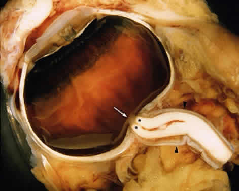
| Fig. 1. Anatomy of the optic nerve head and part of the intraorbital portion of the optic nerve. The intraocular part of the optic nerve consists of the nonmyelinated axons and appears as a semitransparent, yellowish segment (arrow). A portion of the intraorbital optic nerve. The myelinated axons demonstrate a wax-white color (**). The central retinal vessels penetrate into the axia of the optic nerve. The dura mater is continuous with the sclera (arrowhead). |