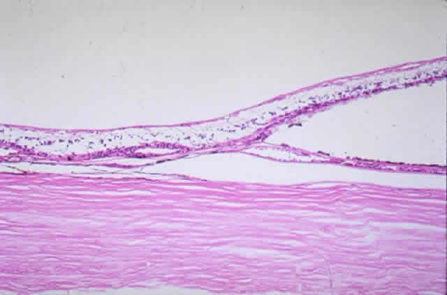
Fig. 33.
Photomicrograph of area corresponding to that in
Figure 31
shows ischemic atrophy of outer retina and retinal pigment epithelium. (H & E, × 25)