|
|
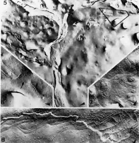
| Fig. 28. Composite of fracture faces of zonulae occludentes between endothelial cells of a choroidal venule. Extensive zonulae occludentes (5) in center. In most places the strands are branching and interconnected; in limited areas they are entirely missing. Higher magnification (6) of delineated area of 5 with interruption (arrow) of the zonula occludens (× 46,000). Typically angulated strands of a zonula occludens (7), seen as imprinted grooves on cell surface (× 42,000). Zonula occludens (8) with cleavage plane jumping from one endothelial cell to another, exposing grooves on cell surface on left and ridges on cell at right (× 54,000). Arrowheads in all figures indicate the direction of platinum-carbon shadowing. (Spitznas M, Reale E: Fracture faces of fenestrations and junctions of endothelial cells in human choroidal vessels. Invest Ophthalmol Vis Sci 14:98, 1975) |