|
|
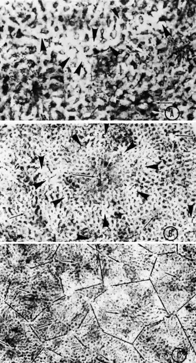
| Fig. 21. Flat preparation of choriocapillaris, posterior pole. A. Arrowheads indicate oval openings to the postcapillary venules. The area around the precapillary arteriole (white-bordered arrow) is stained more heavily because of residual subcapillary collagen. (PAS, × 180; AFIP Neg 74-9984) B. Postcapillary venules (arrowheads) form an irregular ring bordering the capillaries that radiate from the precapillary arteriole (white-bordered arrows), thus outlining a single lobule. The capillaries are broader and clearer near the venules because of less subcapillary collagen. (PAS, × 100, AFIP Neg 74-10240) C. The openings of the postcapillary venules (as shown above) are connected with black lines; they demarcate adjoining lobules in the choriocapillaris. Capillaries from adjoining lobules enter the intervening venules. The lobules form a mosaic of adjoining vascular beds. Precapillary arterioles are indicated by white-bordered arrows. (PAS × 55; AFIP Neg 74-9985) |