|
|
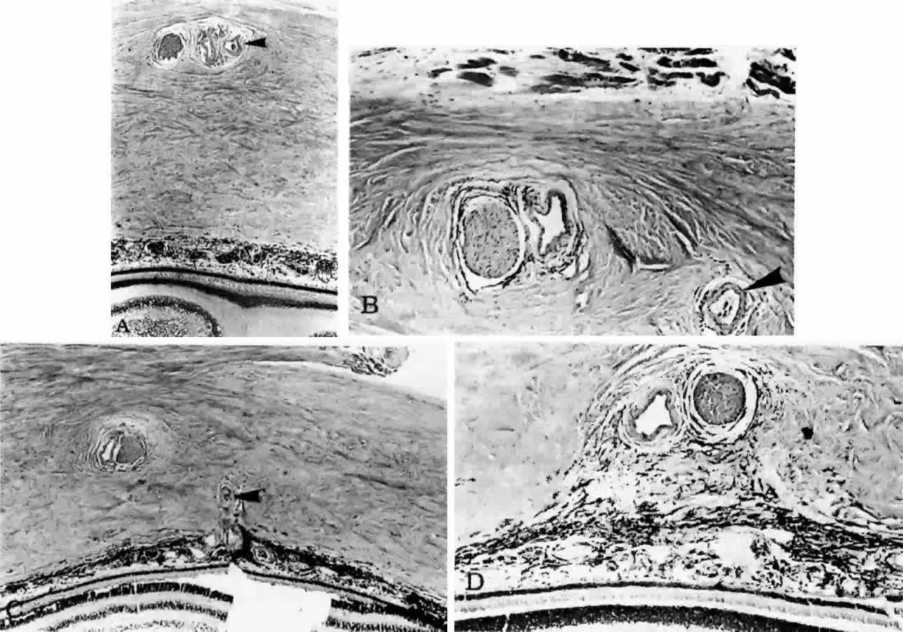
| Fig. 17. Series of tangential sections showing submacular branch of long temporal posterior ciliary artery (LTPCA). The superior pole of the globe (sectioned perpendicular to LTPCA) is on the left; the inferior pole is on the right. A. Foveal region with long temporal posterior ciliary nerve (LTPCN) and LTPCA with small branch (arrowhead). (H & E, × 31) B. LTPCN and LTPCA with submacular branch (arrowhead). Striated fibers of inferior oblique muscle at top. (H & E, × 125) C. Submacular branch (arrowhead) of LTPCA entering choroid. Nerve has now rotated 180° internal to the artery and lies inferior to the artery. Inferior oblique muscle above. (H & E, × 79) D. Melanocytes and fibrocytes in suprachoroid loosely fill in area between LTPCA and LTPCN as they enter the suprachoroid. Faint rim of perivascular-perineural connective cells segregates nerve and artery from sclera. (H & E, × 125) |