|
|
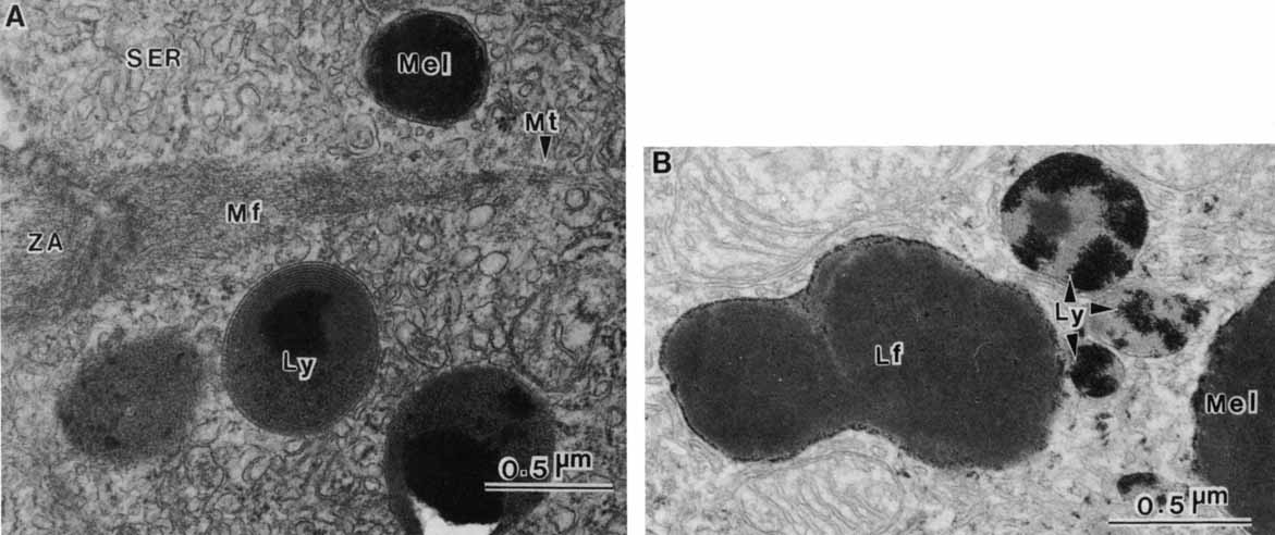
| Fig. 17 Lysosomes. A. Transmission electron micrograph showing a melanosome and three secondary lysosomes near the zonula adherens of infant retinal pigment epithelial cell. The microfilaments (Mf) attached to the zonula adherens (ZA) extend into the cytoplasm amid microtubules (Mt) and the smooth endoplasmic reticulum (SER). A small array of rough endoplasmic reticulum (RER) is also present. The melanosome (Mel) is sectioned across its short axis, so that the cords of melanopolymer are seen in cross section. Note the layer of less dense cortical material. The contents of the lysosomes (Ly) vary in electron density and consistency depending on the identity and the stage of digestion of the contents. Infant retinal pigment epithelium contains no full-size lipofuscin granules; however, very small ultraviolet-fluorescent granules correlate with these secondary lysosomes (×47,500). B. Transmission electron micrograph of retinal pigment epithelium incubated for demonstration of acid phosphatase, a marker lytic enzyme for lysosomes. The lead precipitate formed by the phosphatase activity is localized just inside the membrane of a lipofuscin granule (Lf) and a melanosome (Mel) but is somewhat evenly distributed over the lysosomes (Ly) (×15,000). |