|
|
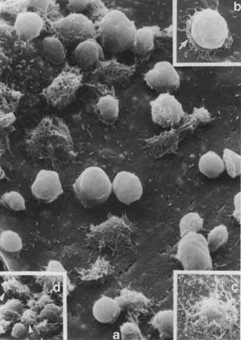
| Fig. 14 Scanning electron micrographs showing the sequence of steps in the phagocytosis of rod outer segment disks by the retinal pigment epithelium. A. An overview of the apical surface of a retinal pigment epithelial cell studded with rod outer segment disks in all stages of engulfment (×4700). B. The first step in phagocytosis involves formation of an attachment “saucer” with a fringe of microvilli (×6500). C. The microvilli then cover the rod outer segment disks (×3850). D. The disk disappears beneath the surface and into the cell. (Chaitin MH, Hall MO: Defective ingestion of rod outer segments by cultured dystrophic rat pigment epithelial cells. Invest Ophthalmol Vis Sci 24:821, 1983) |