|
|
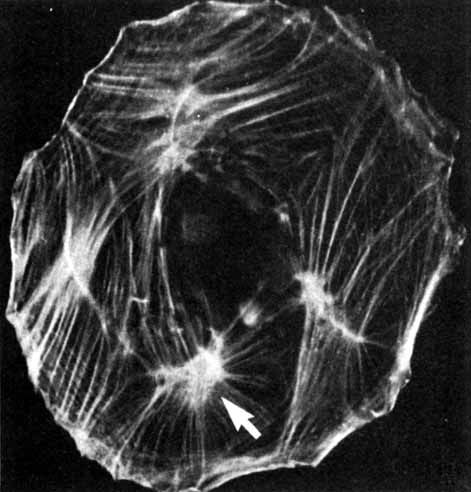
| Fig. 11 Actin microfilament distribution, visualized by fluorescein-labeled antibodies, in a spread preparation of tissue cultured rat retinal pigment epithelium. Actin fibers form parallel arrays (stress fibers) that run along the cell margins and cross the cell, converging at foci (arrow) (×435). (Chaitin MH, Hall MO: Defective ingestion of rod outer segments by cultured dystrophic rat pigment epithelial cells. Invest Ophthalmol Vis Sci 24:821, 1983) |