|
|
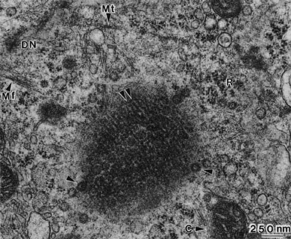
| Fig. 10 High magnification electron micrograph of a grazing section of the retinal pigment epithelial nucleus showing the periodic striations of the chromatin (arrows), nuclear pores (arrowheads), and nuclear-cytoskeletal relationships. Microtubules (Mt), cut length-wise, are slightly larger than ribosomes (R) and approximately the same dimension as mitochondrial cristae (C). Microfilaments (Mf) course in all directions, often as bundles that converge at dense nodes (DN). This specimen came from a 47-year-old patient (×38,000). |