|
|
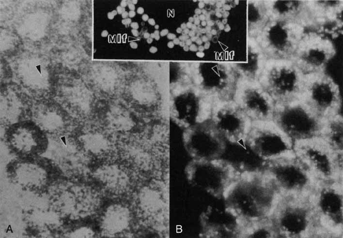
| Fig. 8 Light micrographs of a flat preparation of retinal pigment epithelium from a 22-year-old donor eye. A. By transmitted light retinal pigment epithelial cells appear to be full, except for the area of the nucleus, of opaque brown particles as well as shiny translucent particles. B. When the same specimen is excited by ultraviolet light and the emitted light is properly filtered (i.e., excitation approximately 365 nm, emission >440 nm), the autofluorescence (golden yellow) of the previously translucent lipofuscin granules becomes apparent. Compare granules at arrows (×900). Inset. Fluorescence micrograph of part of a retinal pigment epithelial cell of a 69-year-old donor. Lipofuscin granules are 1 to 1.5 μm in diameter. Complex granules consisting of a central core of melanin and a cortex of fluorescent material are identified, by electron microscopy, as melanolipofuscin granules (Mlf) (×2000). Compare with Figure 12A (N, nucleus) (Feeney L: Lipofuscin and melanin of human retinal pigment epithelium. Invest Ophthalmol Vis Sci 17:583, 1978). |