|
|
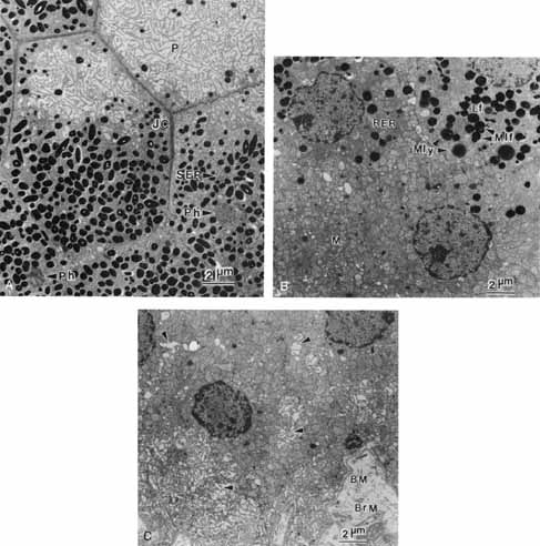
| Fig. 7 Transmission electron micrographs of three successive flat sections through the retinal pigment epithelium of a 26-year-old man (×5600). A. Portions of seven retinal pigment epithelial cells cut across their apical poles. The apical processes (P), forming ridges and microvilli, are shown to better advantage here, than in transverse sections. Profiles of melanin granules in apical processes vary in size depending on the level of the section through the elliptical granule. The hexagonal shape of the cells is visually enhanced by the concentration of various cytoskeletal elements in the region of the intercellular junctional complex (JC). Most of the pigment at this level is melanin. Phagosomes (Ph) and rough endoplasmic reticulum (RER) also lie in the apical pole of the cell. B. Profiles of six retinal pigment epithelial cells sectioned through their middle, at which level the intercellular space is poorly visible. The pigments in this region are principally lipofuscin granules (Lf) and complex granules (arrows), which are large, round melanin granules encased in either lipofuscin or lysosomal enzymes (melanolipofuscin [Mlf]; melanolysosomes [Mly]). The cytoplasm is packed with smooth endoplasmic reticulum (SER) and mitochondria (M). C. Portions of five retinal pigment epithelial cells sectioned across the basal pole where mitochondria are concentrated. The basolateral intercellular space is more prominent (arrows), and it merges with the spaces created by the folded basal plasma membrane. The basement membrane (BM) merges with the inner collagenous zone of Bruch's membrane (BrM). |