|
|
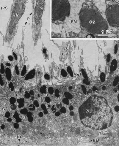
| Fig. 6 Transmission electron micrograph of retinal pigment epithelium in a transverse plane. The basally located nucleus contains a nucleolus (Nu), loose euchromatin and marginated dense heterochromatin. The basal surface of the cell, marked by many infoldings of the plasma membrane, rests on a basement membrane (BM). The apical surface has long processes (P) extending into the interphotoreceptor space (IPS). These processes envelope the distal part of photoreceptor outer segments (OS). The junctional complex (TJ) is the site of attachment between adjacent epithelial cells (arrows). The retinal pigment epithelial cell contains two classes of pigments: elliptical melanin granules (Mel), located apically, and round or figure-eight lipofuscin granules (Lf), located more basally. Phagolysosomes (Ph) and other secondary lysosomes are seen. The cytoplasm contains principally smooth endoplasmic reticulum and a few lamellar arrays of rough endoplasmic reticulum (RER), which is usually located in the apical region. The paranuclear Golgi apparatus (G) is generally inconspicuous. Numerous mitochondria, free ribosomes, and microperoxisomes lie near the infoldings of the basal plasma membrane. Focal adhesions (FA) are special sites connecting the cell to its basement membrane. This specimen is from a 31-year-old woman (×10,500). Inset. Cross-section of outer segments (OS) near the apex of a retinal pigment epithelial cell showing the encircling apical process (P). Note the presence of interphotoreceptor matrix (IPM) in this specimen from a 26-year-old man (×23,000). |