|
|
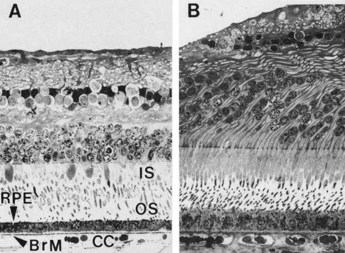
| Fig. 5 Light micrographs of two areas of the same human retina sectioned in a transverse plane and aligned at Bruch's membrane (BrM), demonstrating topographic variation. A. Equatorial retina. Cuboidal retinal pigment epithelial (RPE) cells, with basally located nuclei, form a continuous monolayer that marks the outermost limit of the retina. Each cell extends apical processes into the interphotoreceptor space, which interdigitate with the apical processes (the inner and outer segments [IS, OS]) of the photoreceptor cells of the inverted neurosensory retina. The basal surface of the RPE rests on Bruch's membrane. Capillaries of the choroid (choriocapillaris, [CC]) abut the outer side of Bruch's membrane. B. Fovea. Retinal pigment epithelial cells are taller and narrower. Bruch's membrane is thicker (×320). |