|
|
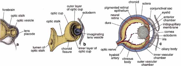
| Fig. 1 Schematic drawing illustrating the development of the eye. Forebrain and developing optic vesicles as seen in a 4-mm embryo (a). Bi-layered optic cup and invaginating lens vesicle as seen in a 7.5-mm embryo (b). The optic stalk connects the developing eye to the brain. The eye as seen in a 15-week fetus (c). All the layers of the eye are established, and the hyaloid artery traverses the vitreous body from the optic disc to the posterior surface of the lens. (Modified from Mann IC, Th Development of the Human Eye. New York, Grune and Stratton, 1974, and reproduced from Ross et al. 2003) |