|
|
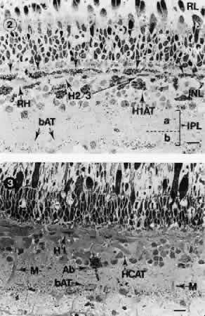
| Fig. 16. Goldfish retina. Light microscope autoradiographs of light-dark differences in the uptake of 3H-γ-aminobutyric acid (GABA). Top. GABA uptake by H1 horizontal cells denoted by downward-pointing arrows just below the outer nuclear layer. Uptake is induced by flickering green light. The axon terminals of H1 horizontal cells (H1AT) also label heavily. Other horizontal cells (H2/3) are unlabeled. bAT, bipolar axon terminals; INL, inner nuclear layer; IPL, inner plexiform layer; a, “off” portion; b, “on” portion; RH, rod horizontal cell; RL, receptor layer. Bottom. GABA uptake in darkness. No uptake is seen by horizontal cell bodies or axons. The Ab-type pyriform amacrine cells (Ab) do label under these conditions, however. HCAT, horizontal cell axon terminal; M, Müller fiber. Marker bars are 10 μm. (Marc RE, Stell WK, Bok D, Lam DM-K: GABA-ergic pathways in the goldfish retina. J Comp Neurol 182:221, 1978) |