|
|
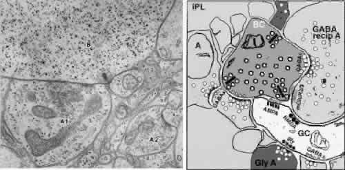
| Fig. 15. Synaptic organization of the inner plexiform layer. The left side is an electron micrograph of a bipolar ribbon synapse in the inner plexiform layer of the carp retina. An amacrine process (A1) receiving input from the bipolar cell (B) makes a reciprocal synapse back onto the bipolar cell terminal. A second amacrine process (A2) synapses onto an unidentified process (× 25,000; data of P. Witkovsky and J. Dowling). The right side is a schematic of the synaptic arrangement at left with the addition of the different neurotransmitter receptor types participating in information exchange. (Courtesy of Dr. Helga Kolb) |