|
|
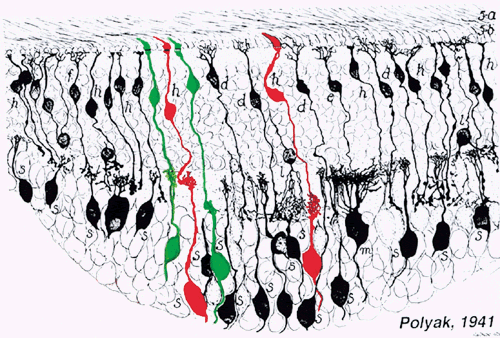
| Fig. 11. The midget pathways of the primate retina. The drawing in black is taken from Polyak S: The Retina. Chicago, University of Chicago Press, 1941. The cells drawn in red and green are midget bipolar cells and midget ganglion cells connecting to L and M cones, respectively. The cone synaptic bases are indicated at top. Each cone type connects to two midget bipolar cells, one of which makes invaginating contacts and ends in the proximal portion of the inner plexiform layer; the other makes basal contacts with cones and ends in the distal portion of the inner plexiform layer. In functional terms, these are, respectively, ON- and OFF-center bipolar cells, which contact ON- and OFF-center midget ganglion cells. (Drawing courtesy of Dr. Helga Kolb) |