|
|
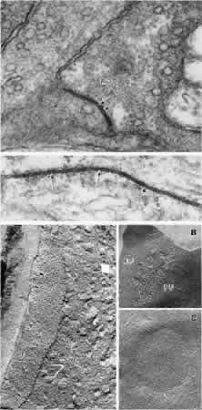
| Fig. 4. Gap junctions in retinal photoreceptors and horizontal cells. Part 1. Gap junctions viewed by transmission electron microscopy. Top. A gap junction (arrows) between a rod base (ROD) and a basal process emitted by a neighboring rod (left) in the Xenopus retina (× 180,000). Bottom. An extensive gap junction (arrows) between two horizontal cell axons in a Xenopus retina (× 216,000; data of P. Witkovsky and C.C. Powell). Part 2. Gap junctions viewed by freeze fracture. A. Gap junction (large arrows) on the protoplasmic face (PF) of the photoreceptor base in the Xenopus retina. At right the receptor cytoplasm is seen in cross fracture; synaptic vesi-cles (SV) are indicated by small arrows (× 109,500). B. Gap junction on the photoreceptor membrane in the Xenopus retina. The fracture plane passed through the junction, revealing both the protoplasmic face (PF), bearing particles, and the external face (EF), bearing pits associated with the gap junction (× 127,750). C. Gap junction on a horizontal cell in the Xenopus retina. The external membrane face contains a plaque of E-face pits (× 27,750; data of A. Nagy and P. Witkovsky). |