|
|
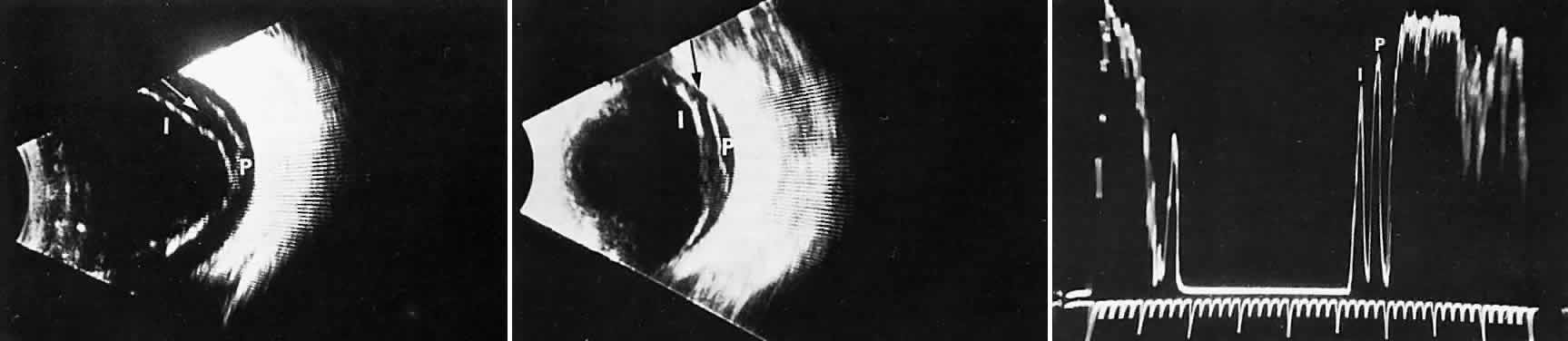
| Fig. 26. Ultrasongraphy of vitreoschisis in the human. Vitreoschisis, splitting of the posterior vitreous cortex (white arrow) can mimic posterior vitreous detachment (PVD). The tissue that remains attached to the macula (P) can induce macular pucker or macular holes. I, inner wall of vitreoschisis cavity; P, outer wall. (From Green RL, Byrne SF. Diagnostic ophthalmic ultrasound. In Ryan SJ, ed. Retina. St. LouisL CV Mosby, 1989, with permission) |