|
|
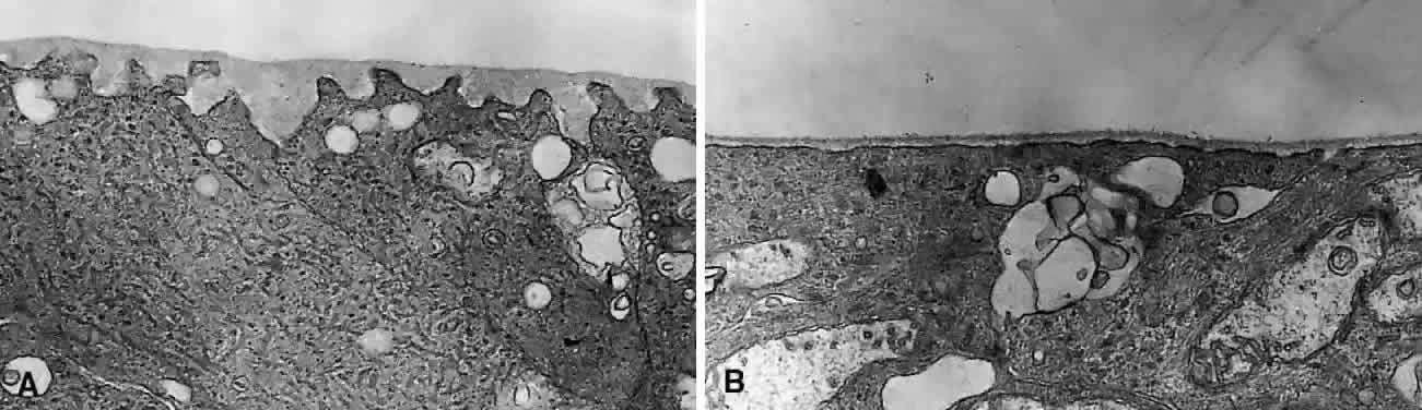
| Fig. 21. Ultrastructure of the human internal limiting lamina (ILL) of the retina. Transmission electron microscopy of the retina from an adult human wherein dissection resulted in a clean separation of retina from the vitreous cortex. The uppermost layer is the ILL with the inner retina below. A: In the posterior pole, the ILL has a smooth anterior surface, whereas the posterior aspect of the ILL is irregular, following the contour of the underlying nerve fibers and Müller cell foot processes. B: In the periphery, both the anterior and posterior aspect of the ILL have a smooth, continuous configuration, without the undulations of the posterior aspect of the ILL that are present in the posterior pole. (Both parts ×4500.) |