|
|
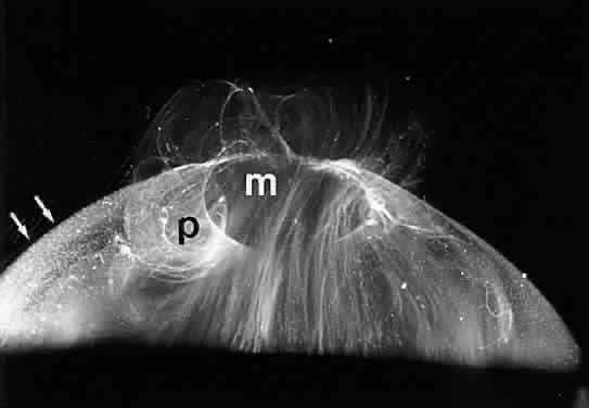
| Fig. 18. Human hyalocytes in the posterior vitreous cortex. Dark-field slit microscopy of dissected human vitreous demonstrating fibers within the corpus vitreus, a hole in the prepapillary posterior vitreous cortex (P), a dehiscence in the premacular vitreous cortex (M), and multiple highly-refractile foci dispersed throughout the vitreous cortex (white arrows). These are hyalocytes. |